Vaginitis

Vaginitis

Vaginitis is an inflammation of the vagina. As many as one third of women will have symptoms of vaginitis sometime during their lives. Vaginitis affects women of all ages but is most common during the reproductive years.


A change in the balance of the yeast and bacteria that normally live in the vagina can result in vaginitis. This causes the lining of the vagina to become inflamed.


Factors that can change the normal balance of the vagina include:


  • Antibiotics
  • Changes in hormone levels due to pregnancy, breastfeeding or menopause
  • Douching
  • Spermicides
  • Sexual Intercourse
  • Infection


The most common types of vaginitis are:


  • Atrophic vaginitis: This condition is not caused by an infection but can cause vaginal discharge and irritation, such as dryness, itching and burning
  • Bacterial vaginosis: This is an overgrowth of the bacteria that occur naturally in the vagina.
  • Trichomoniasis: This condition is caused by the microscopic parasite Trichomonas vaginalis and is spread through sex.
  • Yeast infection: The balance of bacteria and yeast in the vagina is altered, causing the yeast to overgrow and cause symptoms.

What to Expect

To diagnose vaginitis, we will take a sample of the discharge from your vagina and look at it under a microscope. We also may suggest other tests.

Treatment

Treatment will depend on the cause of the vaginitis, and may be either a pill or a cream or gel that is applied to the vagina.

Uveitis

Uveitis

Just as your knee or elbow can become inflamed, so can your eye.  Uveitis is a name for several types of inflammation of the inner part of the eye, often causing impaired vision. It is not a single disease, but a collective term referring to many different conditions within the eye.  '-Itis' means inflammation; therefore, uveitis means inflammation of any part of the uvea (iris, choroid or ciliary body). Uveitis can be controlled with early treatment, but if left untreated may cause serious problems and sight loss. There are different types of uveitis and there can be many different causes. It is very important to know which type you have.  This is because the symptoms, causes and treatment may be completely different.  There are many possible causes of uveitis, such as autoimmune disease, infection, trauma, surgery, or an associated medical condition (such as ulcerative colitis or sarcoidosis).  Often, there is no underlying cause, but a workup may still be done to rule out any systemic causes.

Symptoms

  • Pain in or around the eye
  •  Eye Redness
  •  Sensitivity to light
  • Blurring of vision
  •  Many new floaters
  • Impairment of vision
  • “Pink Eye” that doesn’t get better

 Uterine Fibroids

 Uterine Fibroids

Uterine fibroids are benign (not cancerous) growths that develop from the muscle tissue of the uterus. They also are called leiomyomas or myomas. The size, shape and location of fibroids can vary greatly. They may be present inside the uterus, on its outer surface or within its wall, or attached to it by a stem-like structure. A woman may have only one fibroid or many of varying sizes. A fibroid may remain very small for a long time and suddenly grow rapidly or grow slowly over a number of years.


Fibroids are most common in women aged 30–40 years, but they can occur at any age. Fibroids occur more often in African American women than in white women. They also seem to occur at a younger age and grow more quickly in African American women.

Symptoms

Signs and symptoms of uterine fibroids include:


  • Changes in menstruation
  • Pain during sex or in the abdomen or lower back
  • Pressure, difficulty urinating or frequent urination
  • Constipation, rectal pain, or difficult bowel movements
  • Enlarged uterus and abdomen
  • Miscarriages
  • Infertility


Fibroids also may cause no symptoms at all.

What to Expect

The first signs of fibroids may be detected during a routine pelvic exam. A number of tests may show more information about fibroids:


  • Ultrasonography uses sound waves to create a picture of the uterus and other pelvic organs.
  • Hysteroscopy uses a slender device to see the inside of the uterus. It is inserted through the vagina and cervix (opening of the uterus). This permits us to see fibroids inside the uterine cavity.
  • Hysterosalpingography is a special X-ray test. It may detect abnormal changes in the size and shape of the uterus and fallopian tubes.
  • Sonohysterography is a test in which fluid is put into the uterus through the cervix. Ultrasonography is then used to show the inside of the uterus. The fluid provides a clear picture of the uterine lining.
  • Laparoscopy uses a slender device to help us see the inside of the abdomen. It is inserted through a small cut just below or through the navel. We can see fibroids on the outside of the uterus with the laparoscope.

Treatments

Fibroids that do not cause symptoms, are small or occur in a woman who is nearing menopause often do not require treatment. Certain signs and symptoms may signal the need for treatment:


  • Heavy or painful menstrual periods that cause anemia or that disrupt a woman’s normal activities
  • Bleeding between periods
  • Uncertainty whether the growth is a fibroid or another type of tumor, such as an ovarian tumor
  • Rapid increase in growth of the fibroid
  • Infertility
  • Pelvic pain
  •  

Drug therapy is an option for some women with fibroids. Medications may reduce the heavy bleeding and painful periods that fibroids sometimes cause, but may not prevent the growth of fibroids. Surgery often is needed later. Drug treatment for fibroids includes hormonal birth control, gonadotropin-releasing hormone (GnRH) agonists, and a progestin–releasing intrauterine device.


Myomectomy is the surgical removal of fibroids while leaving the uterus in place. Because a woman keeps her uterus, she may still be able to have children. Fibroids do not regrow after surgery, but new fibroids may develop. If they do, more surgery may be needed. Hysterectomy, the removal of the uterus and possibly the ovaries, is done when other treatments have not worked or are not possible or the fibroids are very large. A woman is no longer able to have children after having a hysterectomy.


Other treatment options include:


  • Hysteroscopy
  • Endometrial ablation
  • Uterine artery embolization (UAE)
  • Magnetic resonance imaging-guided ultrasound surgery

Uterine Cancer

Uterine Cancer

There are two types of uterine cancer: endometrial cancer and uterine sarcoma. Symptoms can include bleeding after menopause, or between periods, and treatment often consists of a hysterectomy, or removal of the uterus.


Endometrial cancer develops in the inner lining of your uterus, or endometrium. Endometrial cancer is one of the most common of the gynecologic cancers.


Uterine sarcoma develops in the muscle wall of your uterus, or the myometrium, and is rare.

Risk Factors

There are multiple risk factors for developing endometrial cancer, and can include an imbalance between estrogen and progesterone, obesity, age, diet, family history, menstrual and reproductive history, and health conditions such as diabetes. Additionally, treatments for other conditions such as radiation therapy to the pelvis and estrogen replacement therapy without progesterone increase your risk of developing uterine cancer.


If you are at high risk for developing uterine cancers, talk to your doctor about steps and lifestyle changes you can make to preserve your health.

Symptoms

Symptoms of endometrial cancer or uterine sarcoma can include:


  • Lower abdominal pain or cramping in the pelvic area
  • Vaginal bleeding between periods
  • Vaginal bleeding or spotting post menopause; or a thin white or clear vaginal discharge
  • Heavy, frequent, or prolonged vaginal bleeding in women over 40

Diagnosis

It is important to talk with your doctor if you experience signs of uterine cancer. Your doctor will review your symptoms, family history and risk factors, in addition to a physical and pelvic exam.


Your doctor may order tests to determine a diagnosis of uterine cancer, including lab tests and imaging tests, such as an MRI, CT scan or transvaginal ultrasound.

Treatments

While your treatment plan will vary based on your overall help and the type and severity of the cancer you have, most people with endometrial cancer require surgery. Most people end up having a hysterectomy, where a surgeon will remove the uterus and the cervix.


Other common treatments may include:


  • Chemotherapy
  • Radiation therapy
  • Hormone therapy
  • Immunotherapy

Prognosis/Life after uterine cancer treatment

Early detection and treatment are key to surviving uterine cancer. Patients are able to return to their normal activities within a reasonable amount of time

Urology Services

Urology Services

At LifeBridge Health, our board-certified urologists provide the latest urology procedures to treat disorders of the urinary tract and male and female urogenital systems involving the bladder, kidneys, ureters, prostate, testicles and penis. We also provide the latest treatment for cancers involving these organs.


Many urologic conditions can be treated with newer medications or minimally invasive procedures. Some of our cost common procedures include, but aren’t limited to:


InterStim® Therapy

A new, minimally invasive procedure that helps control urinary problems through a device implanted under the skin. The device, which is like a “pacemaker for the bladder,” sends a mild electronic impulse to the nerves that control the bladder, sphincter and pelvic floor muscles.


Surgical treatment for urinary incontinence

When other treatments are not working, surgery may be used to support a weakened bladder or pelvic muscles to help you better control urine flow. Procedures include:

  • Repair of pelvic prolapse: urologists can repair prolapsed (sagging) organs through a variety of techniques using meshes and grafts to support the vaginal structures.
  • Sling procedures: minimally invasive technique where your own tissue or synthetic materials are inserted to support your urethra and the neck of the bladder.


Cystectomy

Removal of a part or all of the bladder to treat bladder cancer that has invaded the wall of the bladder. Part of the urethra, nearby lymph nodes and organs (such as the prostate in men or the uterus, cervix and ovaries in women) may be removed in a total (radical) cystectomy. Your surgeon will devise a new internal or external way to pass urine after the surgery.


Extracorporeal Shock Wave Lithotripsy (ESWL)

This non-surgical procedure uses high energy sound waves to break up the kidney stone into smaller pieces that can more easily pass through the urinary tract.


Percutaneous Nephrolithotomy

A tiny incision in the back to remove medium to large kidney stones (renal calculi) from the urinary tract when other treatments are not effective. The stones are removed using a narrow tube inserted through the incision.


Nephrectomy and partial nephrectomy

The removal of part or all of a kidney, often in patients with polycystic kidney disease (multiple cysts), severe kidney infection or kidney cancer, or when a kidney is being donated.


Circumcisions

Removal of the penis foreskin.


Hydrocele repairs

The repair of a fluid filled sac that accumulates in the scrotum, usually benign (not cancerous). It usually involves an outpatient procedure in which the surgeon makes a tiny incision to drain the fluid.


Undescended testicle (cryptorchidism)

Surgery that manipulates the testicle into place in the scrotum, to reduce the increased risk of testicular cancer and infertility that accompanies this condition, which is more common in premature infants.


Minimally Invasive Treatments for BPH (Benign Prostatic Hyperplasia)

Many men experience benign (non-cancerous) enlargement (hyperplasia) of their prostate as they age, which can cause urinary problems such as slow flow, difficulty emptying the bladder or urgency and frequency. Our urologists offer state-of-the-art treatments for this common problem that include:

  • Aqualation Therapy: Aquablation therapy is performed by the AquaBeam Robotic System, the first FDA-cleared, surgical robot utilizing automated tissue resection for the treatment of LUTS due to BPH. It combines real-time, multi-dimensional imaging, automated robotic technology, and heat-free waterjet ablation technology for targeted, controlled, and immediate removal of prostate tissue.
  • Gyrus TURP (transurethral resection of the prostate): TURP is considered the best way to treat an enlarged prostate. This advanced approach uses low-voltage energy to vaporize and remove the portion of the prostate that is enlarged and causing your symptoms.
  • Transurethral microwave thermotherapy of the prostate (TUMT): using heat to destroy excess prostate tissue by inserting a microwave antenna through a special catheter in the bladder.
  • Laser prostatectomy: a minimally invasive procedure that removes excess prostate tissue using a laser.


Radical Prostatectomy to treat localized prostate cancer

Removal of the prostate and surrounding tissue to treat cancer. Minimally invasive techniques can be used to reduce pain and recovery time. It typically involves general anesthesia and a hospital stay of several days.


Urology Care

""

Urology Care

Experience High Quality Care You Can Trust 

We Know Urology Disorders and Diseases Don't Go Unnoticed, Let Us Help

LifeBridge Health’s team of board-certified experts diagnose and treat urologic conditions affecting men, women and adolescents. We provide cutting-edge solutions to treat disorders affecting the bladder, kidney, urinary tract and urologic cancers. Whether surgical intervention is your next step or management through non-surgical techniques, we’re here for you.

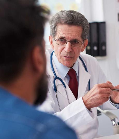
Urology patient with a doctor

Conditions, Treatments and Diagnostics

Experience Advanced Urology Care, Ready For You

LifeBridge Health offers state-of-the-art robotic and minimally invasive surgical options as well as non-surgical therapies options for patients. Our team of clinicians address your individual concerns and offer comprehensive treatment options. Our urologists are here to help you regain control. 

Urology doctor appointment
Teaser Title
Experience High Quality Care You Can Trust 

Urinary Tract Infections

Urinary Tract Infections

Most urinary tract infections (UTIs) start in the lower urinary tract, which is made up of the urethra and bladder. Bacteria from the bowel live on the skin near the anus or in the vagina. These bacteria can spread and enter the urinary tract through the urethra. If they move up the urethra, they may cause a bladder infection (called cystitis). Bacteria that have infected the bladder may travel to the upper urinary tract, the ureters and the kidneys. An infection of the kidneys is called pyelonephritis. An upper urinary tract infection may cause a more severe illness than a lower urinary tract infection.


Women are more likely than men to get UTIs because the urethra is shorter in a woman than in a man. In women, the bacteria can reach the bladder more easily.

Symptoms

One sign is a strong urge to urinate that cannot be delayed. As urine flows, a sharp pain or burning, called dysuria, is felt in the urethra. The urge to urinate then returns minutes later. Soreness may be felt in the lower abdomen, in the back or in the sides.


Other signs may show up in the urine. It may have a strong odor, look cloudy or sometimes be tinged with blood. Blood in the urine also may be caused by other problems. Tell us if you see blood in your urine.


If the bacteria enter the ureters and spread to the kidneys, symptoms also may include back pain, chills, fever, nausea and vomiting. If you have any of these symptoms, tell us right away. Kidney infections are serious and must be treated promptly.

Diagnostics

We will first do a simple test, called urinalysis, to find out whether you have a UTI. For this test, you will be asked to provide a urine sample.

Treatments

Antibiotics are used to treat UTIs. Treatment is usually quick and effective. Most symptoms go away in one to two days. Be sure to take all the medication even though your symptoms may go away before you finish your prescription. If you stop early, the infection may still be present or could come back.

Urinary Incontinence

Urinary Incontinence

Urinary incontinence is a condition that causes an individual to leak urine. Incontinence can range from leaking just a few drops to complete emptying of the bladder.


There are three main types of urinary incontinence:


  • Stress urinary incontinence (SUI) is leaking urine when coughing, laughing or sneezing. Leaks also can happen when a woman walks, runs or exercises.
  • Urgency urinary incontinence is a sudden strong urge to urinate that is hard to stop. Women with this type of urinary incontinence may leak urine on the way to the bathroom. If you have an “overactive bladder,” it means that you have symptoms of urgency and frequency that may or may not include incontinence.
  • Urgency urinary incontinence combines symptoms of both SUI and urgency urinary incontinence.

Symptoms

Signs and symptoms of urinary incontinence include urgency, frequency, waking from sleep to urinate, painful urination, and leaking urine while sleeping.

Diagnostics

The first two steps in assessing urinary incontinence usually are a medical history and physical exam. Sometimes, imaging tests and bladder function tests are done if more information is needed.

Treatments

Nonsurgical treatment options include lifestyle changes, bladder training, physical therapy and using bladder support devices. For urgency urinary incontinence, the treatment may involve medication. Surgery may help certain types of incontinence. Often, several treatments are used together for the best effect.

Urgent Care

""

Urgent Care

When care can't wait

Making Urgent Care Connections

LifeBridge Health partners with Expresscare and Children’s Urgent Care to ensure that our patients have convenient, integrated options for treatment when unable to see their physician. Partnering with these regional leaders in urgent care allows our patients to easily access follow-up treatment at LifeBridge Health facilities.


Patient coming into ExpressCare urgent care in Baltimore for care

ExpressCare

With dozens of convenient locations across Baltimore, Harford, Cecil, Carroll, Anne Arundel and Prince George’s counties, ExpressCare offers pediatric and adult patients an easy alternative to the emergency room for coughs, colds, broken bones, minor cuts and burns and more!

ExpressCare urgent care visit near me

Children’s Urgent Care

Pediatric urgent care is available at all our ExpressCare locations and at Children’s Urgent Care Pediatric Specialists, where child health experts offer care uniquely catered to the needs of children, including strep tests, sports physicals and flu shots as well as treatment for broken bones, colds, infections and more — all in a comforting, kid-friendly environment.

Pediatric Urgent Care Visit in Maryland

Related Blogs and News Releases

Teaser Title
When care can't wait

Ultrasound Exams

Ultrasound Exams

Ultrasound produces pictures of the inside of the body using sound waves. Ultrasound imaging, also called sonography, involves the use of a small transducer and ultrasound gel placed directly on the skin. High-frequency sound waves are transmitted from the probe through the gel into the body. The transducer collects the sounds that bounce back and a computer then uses those sound waves to create an image. Ultrasound exams do not use ionizing radiation, thus there is no radiation exposure to the patient. The procedure may be used to diagnose a variety of conditions, assess organ damage, monitor pregnancy, evaluate pain, swelling and infection, guide procedures like needle biopsies, and much more.


Before the Procedure

You should wear comfortable, loose-fitting clothing for your ultrasound exam. You may need to remove all clothing and jewelry and wear a gown during the procedure. Preparation for the procedure will depend on the type of exam.


During the Procedure

For most ultrasound exams, you will be positioned lying face-up on an exam table that can be tilted or moved. Patients may be turned to either side or on occasion placed in a face-down position to improve the quality of the images. The radiologist or sonographer will apply a warm water-based gel to the area of the body being studied. The gel will help the transducer make secure contact with the body and eliminate air pockets between the transducer and the skin that can block sound waves from passing into your body. In some ultrasound studies, the transducer is attached to a probe and inserted into a natural opening in the body, like the esophagus, rectum or vagina. Most ultrasounds are completed within 30 minutes, although more extensive exams may take up to an hour.


After the Procedure

When the exam is complete, you will be asked to wait until the technologist verifies that the images are of high enough quality for accurate interpretation. After the images have been interpreted, your radiologist or referring physician will discuss the findings with you. Depending on the results of the ultrasound, additional tests or procedures may be scheduled to gather further diagnostic information.

Subscribe to